Fibers, collagen, and fibrosis are emerging biomarkers for oncology
The search for novel biomarkers for oncology and fibrosis has led to increased interest in studying fibers, particularly fibrosis and collagen fibers, because these structural elements of the tumor microenvironment (TME) play significant roles in cancer progression, metastasis, immune response, and drug resistance.
Studying fibrosis and collagen fibers in cancer research is challenging because traditional methods:
- Are labor-intensive
- Require specialized stains and equipment, and access to tissue
- Are challenging to use at scale
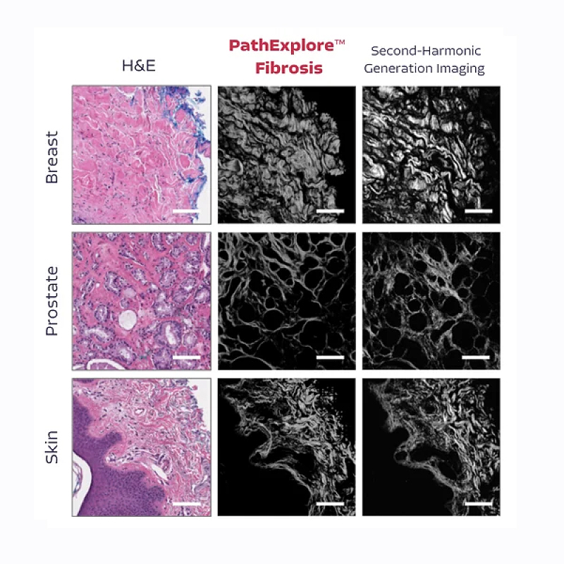
PathExplore™ Fibrosis: Quantify the fibrotic microenvironment
.webp)
Capture and analyze fibrosis, collagen, and fiber directly from routine pathology slides
Fast & Scalable Fibrotic Microenvironment Analysis
Unlike traditional methods that require special stains and complex imaging technology, PathExplore™ Fibrosis leverages AI to quickly analyze routine H&E-stained slides, enabling large-scale studies and fast biomarker discovery.

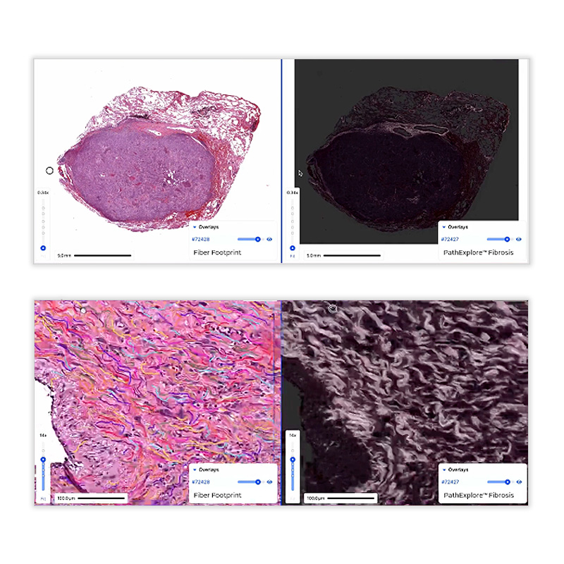
Quantitative Insights
The measurement of fibrosis, collagen, and fiber morphology directly from whole-slide images provides an unprecedented level of detail on tumor microenvironment features, offering new opportunities to explore tumor biology and therapeutic responses.

Integrate with Standard Pathology Workflows
By working with standard pathology workflows (H&E-stained slides), the algorithm democratizes advanced fibrosis analysis, allowing labs to integrate it easily without additional microscopy equipment or complex preparation techniques.
Access PathExplore™ Fibrosis today
Deploy on your slides
- Access PathExplore™ Fibrosis by reaching out at bd@pathai.com or contact us.
See for yourself
- Try our self-serve demo for hands-on experience.
Our Oncology content library


Publication collections


Improving Patient Outcomes with AI-Powered Pathology
Connect with our business development team to learn more about PathExplore Fibrosis™.