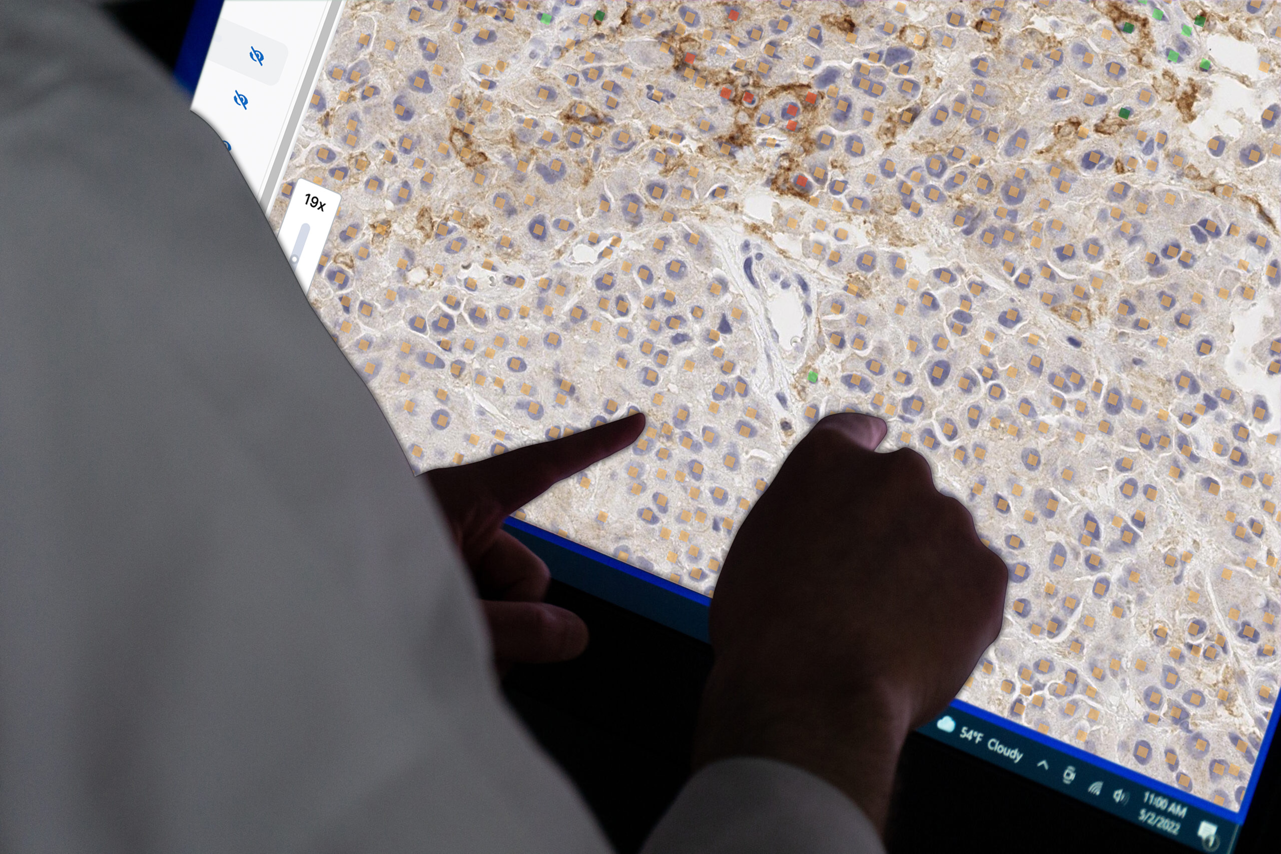
Detect and quantify PD-L1 positive tumor and immune cells while optimizing your digital pathology workflow.

Improve Scoring Confidence
Accurate and reproducible PD-L1 scoring. Testing demonstrated high concordance with consensus pathologist review

Streamline Workflows
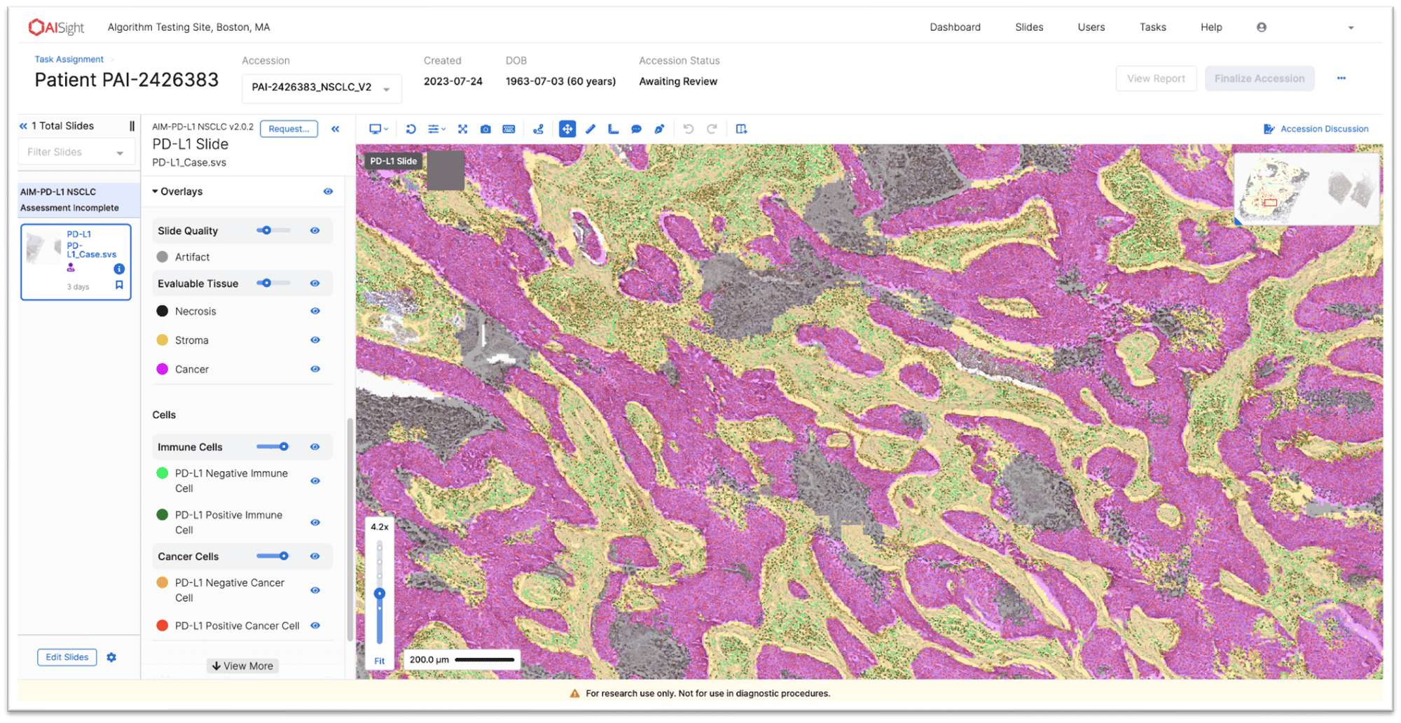
Automated scoring, whole-slide image analysis, and tissue region exclusion (e.g.,control tissue)

Robust Development
Trained on >10,000 samples and >2 million annotations from board-certified pathologists.
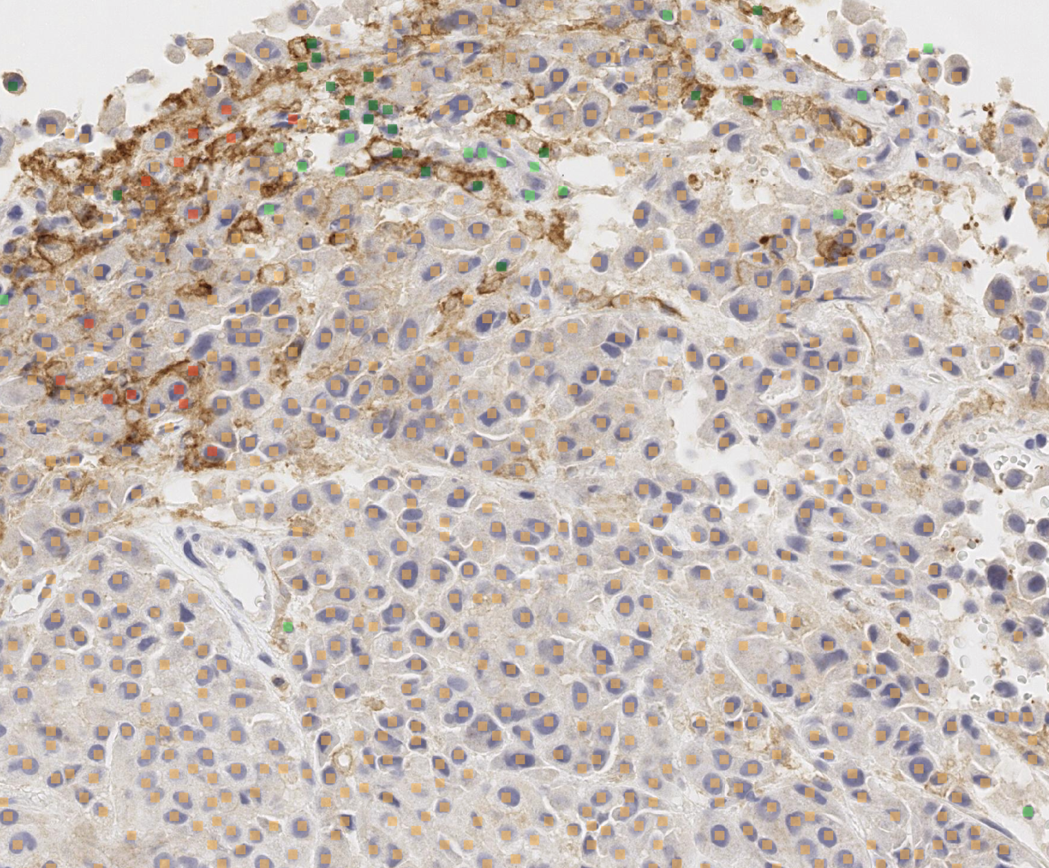
AIM-PD-L1 NSCLC
AIM-PD-L1 NSCLC is multi-clone and multi-scanner compatible
Specifications:
- Intended Use: Research Use Only
- Indications: Non-small Cell Lung Cancer
- Clones: Agilent Dako 28-8 and 22C3; Roche Ventana SP142 and SP263
- Scanners: Leica Aperio® AT2 and GT450; Philips UFS, Roche DP200, Hamamatsu NanoZoomer® s360
- Inputs: Biopsy and/or resection sample from primary, lymph node, or metastatic tumor
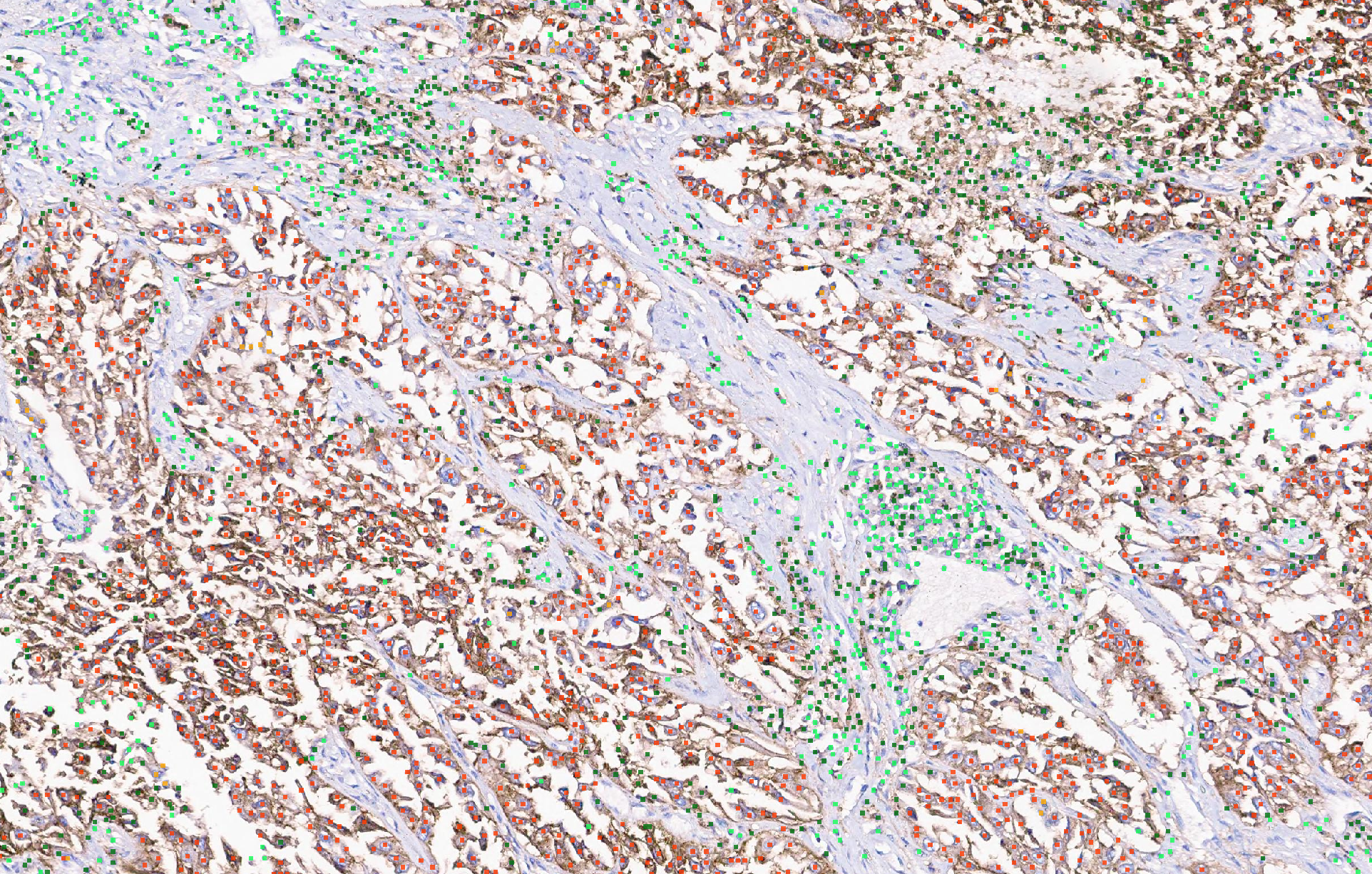
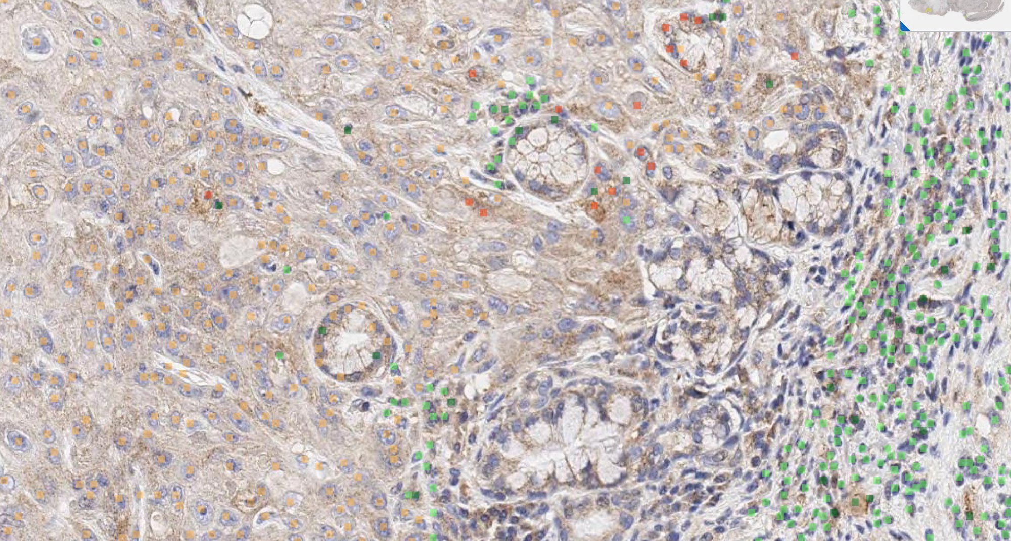
- Outputs: Percent PD-L1 positive tumor cells and proportion of PD-L1+ cells to all cancer cells; cell counts and proportion across cell classes in different tissue regions
AIM-PD-L1 HNSCC
Specifications:
- Intended Use: Research Use Only
- Indications: Head and Neck Squamous Cell Carcinoma
- Clones: Agilent Dako 28-8
- Scanners: Leica Aperio® AT2 and GT450; Philips UFS, Roche DP200, Hamamatsu NanoZoomer® s360
- Inputs: Biopsy and/or resection sample from primary, lymph node, or metastatic tumor
- Outputs: Percent PD-L1 positive tumor cells and proportion of PD-L1+ cells to all cancer cells; cell counts and proportion across cell classes in different tissue regions
AIM-PD-L1 Melanoma
Specifications:
- Intended Use: Research Use Only
- Indications: Melanoma
- Clones: Agilent Dako 28-8
- Scanners: Leica Aperio® AT2 and GT450; Philips UFS, Roche DP200, Hamamatsu NanoZoomer® s360
- Inputs: Biopsy and/or resection sample from primary, lymph node, or metastatic tumor
- Outputs: Percent PD-L1 positive tumor cells and proportion of PD-L1+ cells to all cancer cells; cell counts and proportion across cell classes in different tissue regions
AIM-PD-L1 UC
Specifications:
- Intended Use: Research Use Only
- Indications: Urothelial Carcinoma
- Clones: Agilent Dako 28-8
- Scanners: Leica Aperio® AT2 and GT450; Philips UFS, Roche DP200, Hamamatsu NanoZoomer® s360
- Inputs: Biopsy and/or resection sample from primary, lymph node, or metastatic tumor
- Outputs: Percent PD-L1 positive tumor cells and proportion of PD-L1+ cells to all cancer cells; cell counts and proportion across cell classes in different tissue regions
.webp?width=1746&height=1278&name=Screen-Shot-2023-07-26-at-10%20(1).webp)